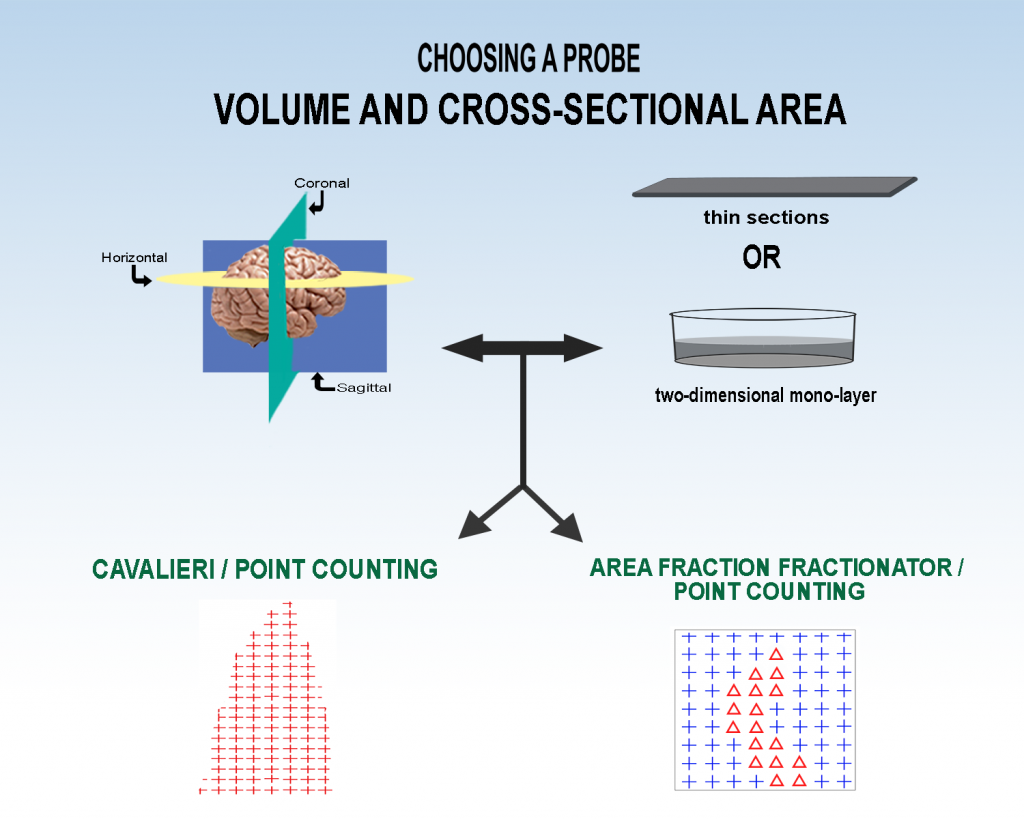
Cavalieri (segmentation)/point-counting
To estimate volume of anatomical regions or components of regions, combine the Cavalieri method of segmentation with point-counting. Using this method to estimate the volumes of particles, however, is problematic because its difficult to resolve their tops and bottoms under the microscope (Tandrup, etal., 1997, pg. 108, last paragraph). The Cavalieri method involves systematic random sampling through the region of interest. Then on the sections that are randomly and systematically chosen, instead of tracing the region, points that are over the region of interest are marked. Each point has an area associated with it; if you think of the points being laid out in a grid with a side length of ‘g’ then the area associated with each point is g*g. the area estimate for a given section is the number of points times their associated area. The volume estimate multiplies this by the thickness and interval of the sections:
For an example of unbiased estimation of volume with the Cavalieri/point-counting method see ‘Cavalieri Estimator‘.
Area Fraction Fractionator
To estimate the volume to volume or area to area percentage of one type of tissue per another type of tissue (for instance lesion or plaque load), systematic random sampling and point counting can also be used, but instead of on the whole region, a fraction of the region can be sampled (Area Fraction Fractionator). A two-dimensional virtual space, usually a square or rectangle, can be superimposed on the region, also using systematic random sampling. An array of points is in each sampling box, and one marker is marked on points of one type of tissue (tissue A) while another marker is used for the other type of tissue (tissue B). The number of points over tissue A (subregion) is divided by the total number of points (reference) to get an estimate of the volume to volume percentage of tissue A per tissue A plus B:
Howard, C.V. and M.G. Reed (2010) Unbiased Stereology, Second Edition, Chapter 4, Estimation of Component Volume and Volume Fraction, QTP Publications, Liverpool, U.K.
García-Fiñana, M., L.M Cruz-Orive, C.E. Mackay, B. Pakkenberg, and N. Roberts (2003) Comparison of MR Imaging Against Physical Sectioning to Estimate the Volume of Human Cerebral Compartments, NeuroImage, 18, 505 – 516.
Tandrup, T., Gundersen, H.J.G. and E.B. Vedel Jensen (1997) The Optical Rotator. J. of Microscopy, 186, 108 -120.
____________________________________________________________________

Sponsored by MBF Bioscience
developers of Stereo Investigator, the world’s most cited stereology system


