The vertical uniform random (or VUR) sections is used for some stereological methods, particularly with tissues that have well defined orientations, such as skin, muscle, and bone. VUR sections are sometimes referred to as vertical sections. A VUR section is less random than a completely random section, the IUR section. A common use of VUR sections is to estimate length and surface area. In some fields, such as Neuroscience, the imposition of VUR sectioning was not accepted as practical. This led to the development of length and surface probes such as the Space Balls and Isotropic Fakir method that could be used in SRS preferential sections. Nevertheless, VUR sections are sometimes appropriate and are described below.
A VUR section or slice is made as such:
- Select an arbitrary vertical axis, or select a horizontal plane and then determine the axis perpendicular to that plane. Either way an orientation is selected that is called the vertical axis.
- Rotate the material in a random manner about the vertical axis.
- Cut sections or slices parallel to the vertical axis that have a random start position.

In Figure 1, a piece of tissue from a biopsy has been mounted in a block. The long axis of the biopsy tissue has been chosen as the vertical axis. In the middle picture, a random rotation angle has been chosen and the rotation has been applied around the vertical axis. In the final step, the tissue is sectioned. Just as an SRS section has a random start, so do vertical sections.
The selection of the vertical axis often is natural because of the nature of the material being studied. Studies of skin have a natural flat surface. This can be considered the horizontal plane. The orientation of a plane can be described by the vector that is perpendicular to the plane. The vertical axis is the orientation that is perpendicular to the plane defined by the surface of the skin.
The selection of the horizontal plane and its associated vertical axis does not have to make any sense relative to the material. For example, a muscle is a highly oriented feature. It is not necessary to have the vertical axis match the long axis of the muscle. What is important is that the vertical axis is maintained throughout the work.

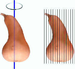
Figure 2 is a butternut squash that has been rotated around a vertical axis. The squash is then sliced to prepare a number of vertical slices. Notice that some of the slicing planes cut the squash to form 2 separate pieces. This occurs on the left side of the squash.
The vertical sections cut from the squash can be used to estimate surface area. Since these sections are more random than the preferential sections that can be used for Cavalieri /point-counting, these same sections can be used for volume estimation.

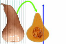
After the slices or sections have been made it is important to know the orientation of the vertical axis on that slice or section. Figure 3 shows the butternut squash again. The vertical slice has been rotated to show the profile of the slice. The green arrow identifies which slice is being shown. The blue arrow identifies the vertical axis. The vertical axis is a direction. The position of the blue arrow is not important. All that matters is the direction that the blue arrow points. This direction is important once probes are placed over the sections or slices that are used. This is because a special line-segment, called a cycloid must be used for the probe and the cycloids must be properly lined up with the vertical direction.
Baddeley AJ, Gundersen HJ, Cruz-Orive LM. J Microsc 1985;142:259.
A method of creating random sections while following an atlas has been described by Dorph-Peterson. This paper describes the detailed laboratory procedures that combine preferential cutting of an organ with the final preparation of random sections. The preferential cutting of the brains was necessary to follow an atlas to the portion of the brain that was being investigated. Once the proper portion of the brain was located, random sections were produced. The sections created were VUR sections, but the same steps can lead to the creation of IUR sections.
Dorph-Petersen KA., J Microsc 1999;195:79.
A vertical section will randomize the tissue in two planes. When the section is under the microscope, the probing lines or planes are randomly thrown down on the tissue and that completes the randomization process. But since a random angle was picked when the tissue was spun about the vertical axis, a degree of freedom has been lost. In other words, the angle that is picked for the orientation of the probes can’t be random it must be sine-weighted. In practice, the shape of the probe can be made sine-weighted instead of picking the angle in a sine-weighted fashion. The appropriate shape is a cycloid.
____________________________________________________________________

Sponsored by MBF Bioscience
developers of Stereo Investigator, the world’s most cited stereology system
