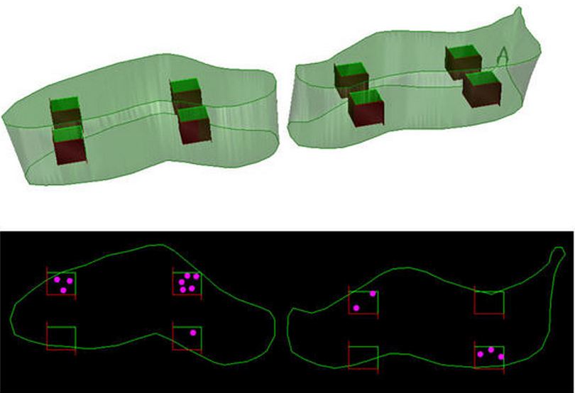
In general, investigations on the number of cells in a region of interest are complicated by the fact that it is usually not possible to physically isolate the cells. Accordingly, it is necessary to cut the tissue into sections, and to find the number of cells by inspecting the sections. This, however, leads to the problem that cutting a given tissue volume into sections also results in cutting the cells in this tissue. Obviously, the number of cell fragments in the sections differs from the original number of cells in the tissue, and as a consequence, estimates of cell numbers based solely on counts of cell fragments in sections are biased (Using Unbiased Stereology to Accurately Determine the Number of Cells). Design-based stereology provides two methods to perform this quantification.
The first uses the fractionator method and is called the Optical Fractionator. It uses thick sections and estimates the total number of cells from the number of cells sampled with a Systematic Randomly Sampled (SRS) set of unbiased virtual counting spaces covering the entire region of interest with uniform distance between unbiased virtual counting spaces in directions X, Y and Z. Another method calculates the mean cell density (Nv) within the unbiased virtual counting spaces by dividing the number of cells counted within all counting spaces by the number of investigated counting spaces and their uniform volume. When multiplying this average cell density with the volume of the investigated region of interest (Vref can be estimated with the Cavalieri estimator), one obtains an unbiased estimate of the total number of cells in the region of interest. This principle has been named the NvVref method. Because no reference volume is required for estimation of number with the Optical Fractionator, it is easier to implement. Additionally, it is easier to estimate the precision of the number estimate (the CE) generated using the Optical Fractionator, making it the method of choice.

The Optical Fractionator is the most commonly used stereological probe in the life sciences. It can be used to estimate the total number of objects in any three dimensional volume regardless of that volume’s shape. The Optical Fractionator is appropriate for the quantification of such objects as cells, synapses, glomeruli, and any other discrete objects within biological tissue. The Optical Fractionator requires that tissue sections be thick for the analysis of neurons. The tissue to be analyzed may be sectioned in any orientation (also known as preferential sectioning). The cell counts carried out with the Optical Fractionator are unbiased in that they are not influenced by the size, shape, spatial orientation, and spatial distribution of the cells under study.
The Optical Fractionator starts by using the fractionator principle to select a series of systematically random sampled (SRS) sections and then sampling each section in the X,Y, and Z axis, again, by using the fractionator principle. By sampling a subset of the total number of objects within the thickness of the tissue and extrapolating, estimates are generated for the total number of particles in the entire region of interest. Estimates generated by the Optical Fractionator may be statistically evaluated.
For more information on the optical fractionator and other length probes please see ‘Number‘.
____________________________________________________________________

Sponsored by MBF Bioscience
developers of Stereo Investigator, the world’s most cited stereology system